Dr. Brian Nagy | January 30th, 2018
Posted In: General Foot Care
 Your feet do more than get you from place to place; they support the weight and joints of your entire body. Keeping your feet healthy helps keep your body strong and healthy, too. So how is it that your feet can stand all that pressure? The answer lies within the structure of the foot itself.
Three Areas of Foot Bones
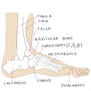
The first thing to know about the foot is to understand the three basic sections of bones. The forefoot is comprised of your toes (phalanges) and the five longer bones (metatarsals) connected to them. The midfoot includes all the bones that make up your arches (navicular, cuboid, and cuneiform). Your hindfoot is your heel (calcaneus) and ankle (talus) bones. If you have an x-ray that reveals trouble in your cuneiform or midfoot, that means you have a problem in the arch of your foot. Easy so far, right?
The 26 bones in each human foot are organized in such a way to form arches, which are like small bridges. Each of your feet has three arches; two running along their length and one that runs across your feet. Your arches help your foot flex when you’re walking on uneven ground so your body remains supported. But arches don’t support themselves; they get a “lift” from your ligaments.
Ligaments are bands of tissue that support your bones and bind them together. Your foot is comprised of various ligaments, but the main ones lie underneath your foot bones. Strong ligaments create a firm yet flexible base that supports the weight of your whole body and helps you move. Your New Hampshire podiatrist may advise you to do exercises that strengthen your ligaments if your feet or ankles are weak. Additionally, certain types of stretches can releive nagging foot pain.
Your foot bones, arches, and ligaments don’t move themselves; that’s what muscles are for. Movement of your foot is caused in large part by muscles in your lower leg. These muscles are attached to your feet by tendons and make your foot muscles more powerful. Muscles on the top of your feet (extensors) allow you to move and stretch your toes. You also have four layers of muscle on the bottoms or soles of your feet.
The first layer is closest to the surface of the sole of your feet and aids in the flexing or bending of your toes. They are somewhat like the muscles in the your hands that help your fingers move, but do not enable quite the same movement your fingers have; most people don’t eat, write, or do laundry with their feet! The other three layers include tendons and although each layer has its own purpose, they mainly work together with the rest of your foot muscles to keep your arches stable. They also help maintain the overall stability and flexibility of your feet.
If you have questions or concerns about the anatomy of your feet, contact Dr. Nagy, your qualified, experienced, and compassionate podiatrist in New Hampshire. At Nagy Footcare, we believe you deserve to be listened to. We believe you deserve to have your questions and concerns addressed. If you are suffering from foot or ankle pain, we believe you deserve to move toward becoming pain free. We believe you should fully understand what is causing your pain and what your best treatment options are. We believe you should have access to the newest technologies and treatments available, and we want to help you. Call Nagy Footcare, your New Hampshire podiatrist, today.
At Nagy Footcare, our best day is when you wake up with no foot pain.
« How to Help Your Foot Pain While on the Job | 45 Percent of People Have This Foot Problem…Do You? »
![]()
PORTSMOUTH
875 Greenland Road
Unit C-4
Portsmouth, NH 03801
(603) 964-6555
(603) 516-4411
FAX: (603) 964-6515

